The Instrument
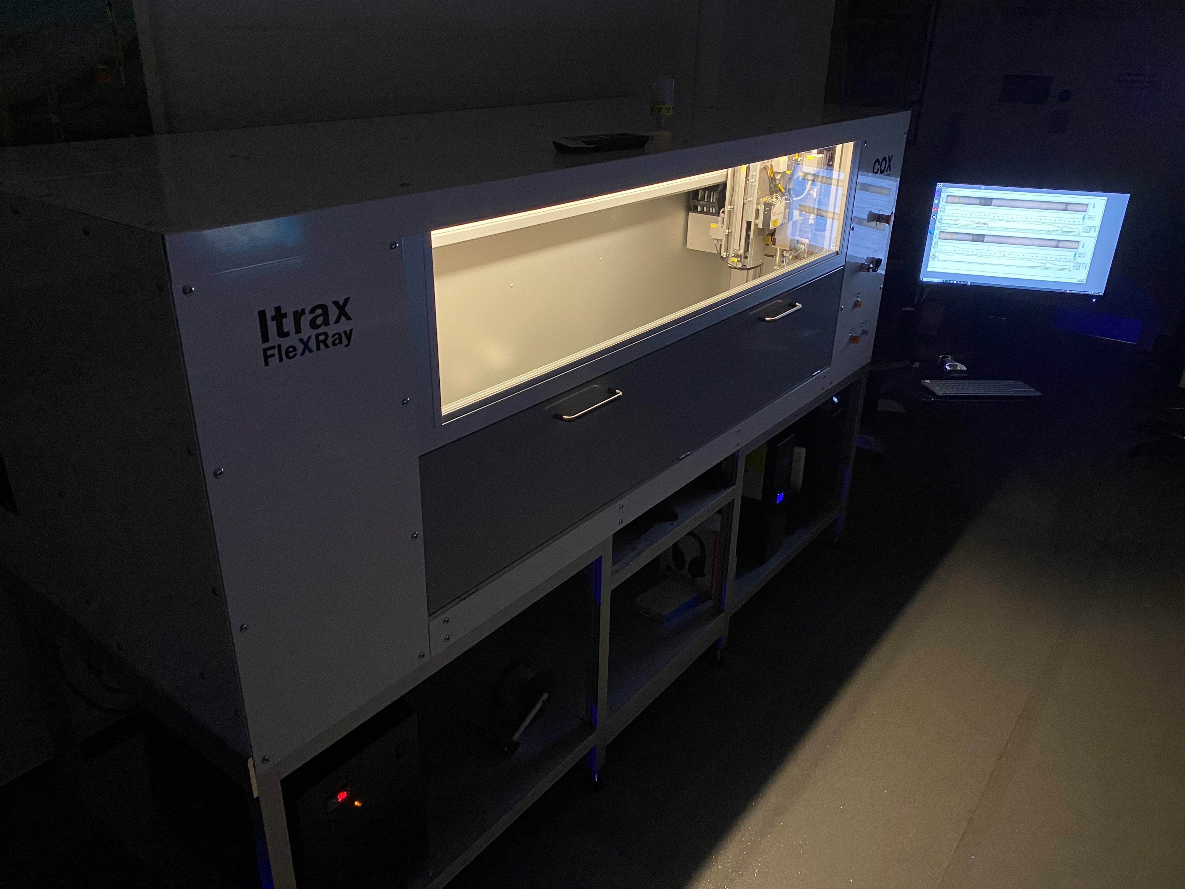
The COX Analytical Systems Itrax X-ray Fluorescence (XRF) Scanner assesses the elemental composition of a sample, most often a sediment core. In addition, the Itrax captures a high-resolution optical sample-surface image and a X-radiograph that assesses whole-sample density contrasts.
Major benefits of analysing samples by Itrax XRF scanning:
- Rapid data collection – large data sets in a timely manner
- Non-destructive – samples remain intact for reproducibility tests, discrete sampling, or archiving
- High resolution – adjustable step-sizes between 200 micrometre and 2 cm
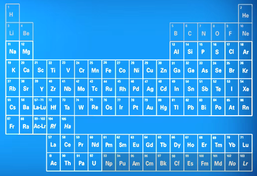
- Study specific – able to target specific elements in the range from Silica to Uranium
This versatility has made the Itrax XRF scanner one of the most widely used analytical instruments in the Earth and Environmental sciences. Further applications of Itrax XRF scanning are found in the Forensic, Material, Archaeological, Biological, and Palaeontological sciences.
Samples and Methods
The Itrax is primarily designed for sediment core samples. However, a broader suite of samples can be run on the Itrax.
Samples successfully run on the Itrax XRF scanner:
- Sediment, peat, and soil samples, such as split-cores and U-channels.
- Rock samples, such as drop stones, nodules, stalagmites, and stalactites.
- Fossils, such as (remineralized) bones, teeth, corals, and shells.
- Other materials, such as wood, glass, ceramics (stone- and earthenware), brick and building materials, and (pre-)historic art objects.
Sample preparation
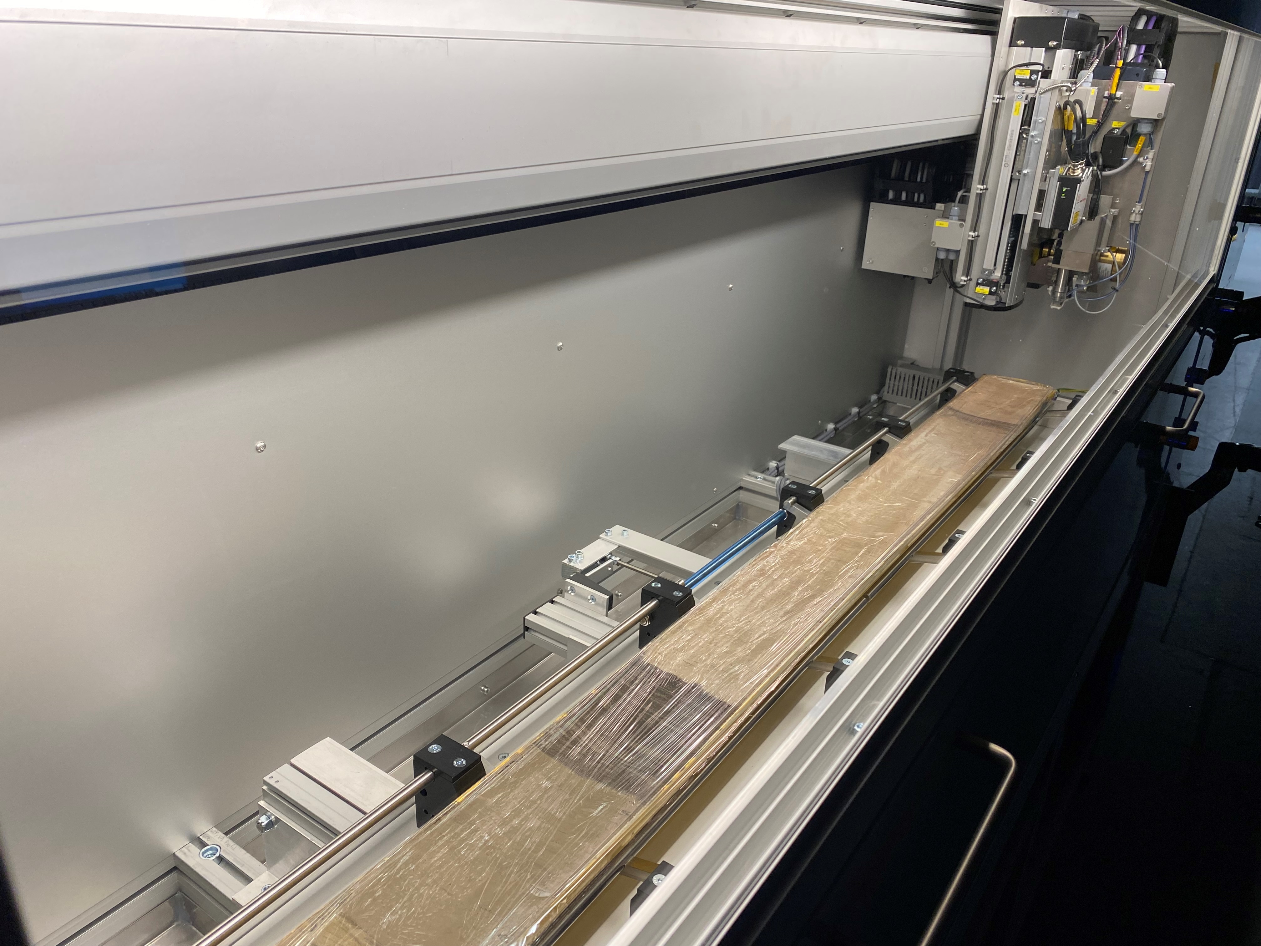
Please note, samples should not be wider than 12 cm, longer than 180 cm, or thinner than 1 cm. A clean and flat sample surface yields optimal results. Therefore, sample preparation, such as splitting and scraping of sediment core samples, or cutting of rock and fossil samples, is needed prior to analysis.
Measurement procedure
After sample preparation, the sample is loaded in the scanner, and an optical image is captured. Instrument settings are selected and optimized for the X-radiograph and XRF scan of the sample. Data acquisition and processing using in-house software can commence.
Data and Applications
The Itrax XRF scanner can collect element profiles in the range from Sodium to Uranium. XRF results are output as peak areas (total counts), which are a relative and semi-quantitative measure of elemental concentrations.
Scientific Applications
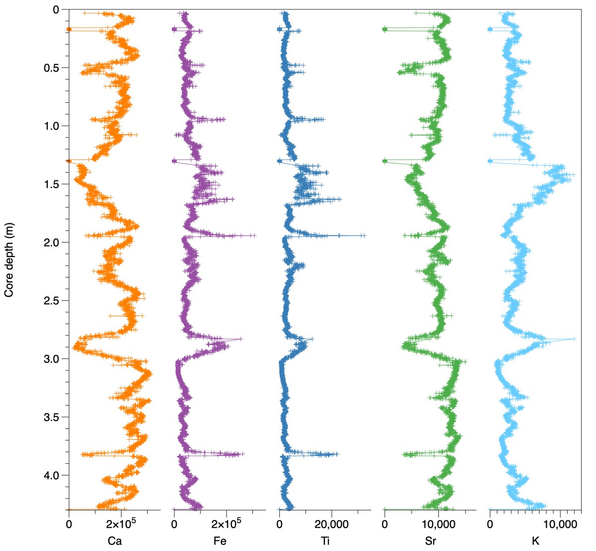
By combining the optical image, X-radiographic, and XRF data, allows for many advanced scientific applications:
- First-order (geo-) chemical characterizations of material make-up.
- Description of environmental change.
- Tracking the distribution of contaminants throughout a core.
- Identification of diagenetic relocation of particular elements, such as the vertical migration of redox fronts in the sediment column.
- Characterisation of palaeosols.
- Identification of bed provenance, for examples layers with volcanic ash, ice-rafted debris, and other event stratigraphies.
- Stratigraphic descriptions and correlation between drill-holes.
- Cyclostratigraphy and astrochronology.
- Sclerochronology and the description of varved (annually layered) sediments.