What is the Geotek XCT System?
The Geotek XCT system at BOSCORF is a state-of-the-art X-ray computed tomography scanner specifically designed for analysing sediment cores, rock cores, soil samples, and other environmental materials. It is like a medical CT scanner, but engineered for geological and environmental samples. It allows for observing materials inside cores without cutting them open, revealing internal structures, layers, and features that would otherwise be invisible or destroyed during traditional analysis.
Advantages of BOSCORF's XCT System
Allows for routine analysis: Unlike many CT systems, this cabinet-based design is fully shielded, meaning you don't need an expensive lead-lined room. The system can be used in any standard laboratory setting. Meaning uninterrupted workflows in the lab.
Ideal for Environmental Research: The system is specifically designed for the types of samples environmental scientists work with, from marine sediment cores to rock cores to permafrost samples to soil columns.
Complete workflow: BOSCORF provides the scanner and the ability to perform the entire workflow from scanning to final analysis, backed by high-performance computing and software resources.
Example Studies
Sediment Cores: Examine layering, bioturbation, and sedimentary structures in cores from lakes, oceans, and rivers without disturbing the sample.
Rock Cores: Analyse geological structures, fracture networks, porosity, and mineral distribution in bedrock samples from drilling programs.
Climate Records: Study seasonal layers (varves), ice content, and climate indicators preserved in sediment.
Soil Structure: Analyse soil architecture, root systems, pore networks, and how they change over time or with different treatments.
Pollution Studies: Track how pollutants move through sediments, rocks, and soils, identifying migration pathways and distribution patterns.
Permafrost Research: Examine ice content, organic matter distribution, and structural changes in frozen ground samples.
Wetland Ecology: Study peat structure, plant remains, and decomposition patterns in wetland cores.
Operation
The system works by rotating your core sample while X-rays pass through it from different angles. A high-resolution detector captures these X-ray images, and computer algorithms reconstruct them into detailed 3D images. You can then "slice" through your sample virtually, examining it from any angle or direction.
Sample Preparation: Minimal preparation is needed. The system can scan whole cores or cut sections. Cores can be 2.5 to 14 centimetres in diameter and up to 1.5 meters long.
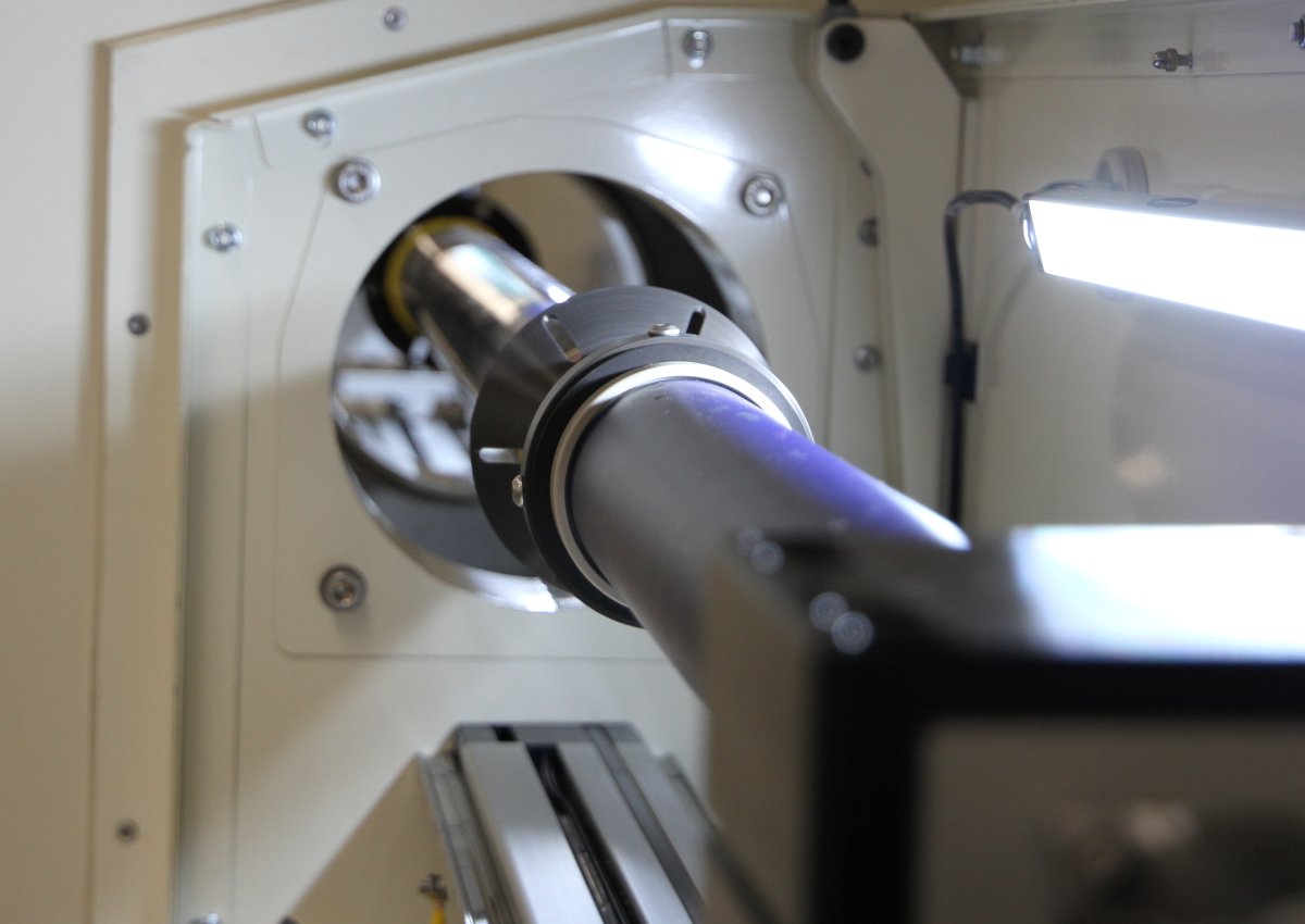
Scanning Process: Samples are loaded horizontally into motorised arms that hold them securely. The system rotates the core while X-rays pass through it, creating thousands of images from different angles.
Image Quality: The system can achieve incredible detail, with resolution ranging from 30 to 350 micrometres (finer than the width of a human hair).
Technical Capabilities and Sample Requirements
Imaging Options:
• 2D Radiography: Quick X-ray images that show internal structure
• 3D Laminography: Focused imaging of specific layers within your sample, ideal for split cores. This means there is no longer a need to slab cores.
• 3D Circular or Helical and start/stop or sync CT: Complete 3D reconstruction showing every detail of your core's internal structure
Notes on Sample Types:
• Full 3D CT scanning is only available for whole cores, the system needs to rotate the complete sample 360 degrees to create the full 3D reconstruction
• For split cores, we recommend laminography, this technique can examine specific layers and structures within split core sections without requiring full rotation
• Both whole and split cores can be analysed using 2D radiography for quick structural assessment
Power Options
The XCT is a 130kV system, ideal for a range of sample types. The flat-panel detector has nearly 3 million pixels (1920 x 1536). The system also comes fitted with a full suite of filters and scanning options.
BOSCORF's Computing Resources
Scanning is just the first step, the real power comes from our high-performance computing capabilities:
Data Processing: Raw CT data requires significant computational power to reconstruct into usable 3D images. Our HPC cluster can handle this processing quickly and efficiently.
Advanced Reconstruction: We use sophisticated algorithms to turn thousands of X-ray projections into detailed 3D volumes you can explore from any angle.
Segmentation: Our computing resources allow the identification of separate materials, layers, or features within your samples.
Quantitative Analysis: Beyond just looking at images, it is possible to measure volumes, densities, porosities, and other quantitative properties throughout your samples.
3D Visualisation: Create publication-ready images, movies, and interactive 3D models of your data.
Software Tools Available
Geotek X-ray Acquisition Software: Control scanning parameters and monitor data quality in real-time.
Geotek Reconstructor: Powerful reconstruction algorithms that turn your raw scan data into 3D volumes or laminographic sections.
Geotek GQuickView: Interactive tools for viewing and manipulating X-ray images and CT slices.
Geotek CTQuickView: Advanced 3D visualization and analysis tools for exploring your reconstructed volumes.
HPC Cluster: Allows efficient visualisation and processing of full sample files in Aviso.
Choosing the Right Technique for Your Samples
Use 3D Helical CT when you have:
• Whole cores in good condition
• Need complete 3D structural information
• Want to measure volumes and 3D connectivity
• Require full quantitative analysis capabilities
Use Laminography when you have:
• Split cores or core sections
• Need detailed examination of specific layers
• Want to focus on particular structural features
Use 2D Radiography when you need:
• Quick structural overview of any core type
• Rapid assessment of multiple samples
• Basic density and structure information
• Preliminary evaluation before detailed scanning
Getting Started
BOSCORF provides comprehensive support throughout your research project. Whether you're new to CT scanning or an experienced user, our team can help you design the best scanning strategy for your samples and research questions.
We'll work with you to determine whether your cores are suitable for full 3D CT or whether laminography would be more appropriate, ensuring you get the best possible data from your valuable samples.
The combination of cutting-edge imaging technology and powerful computational resources makes BOSCORF's XCT system an invaluable tool for research
Contact BOSCORF to discuss how X-ray CT can help your research. Our team will help you choose the right imaging approach based on your core condition and research objectives.